[Feature] Introducing a New Method for Monitoring Blood Vessels in Cardiovascular Disease Prevention
The research team led by Professor Park Bum-jun from the Dept. of Chemical Engineering at Kyung Hee University (KHU) has introduced a new vascular monitoring method that could contribute to the early detection of vascular stenosis. The team used a sensor made from polydiacetylene (PDA) particles, which exhibit fluorescence under mechanical stress, to examine the vascular stenosis model and address limitations in previous research. The study was published in the prominent journal Nature Communications on July 19, 2024, under the title A Single-Particle Mechanofluorescent Sensor.
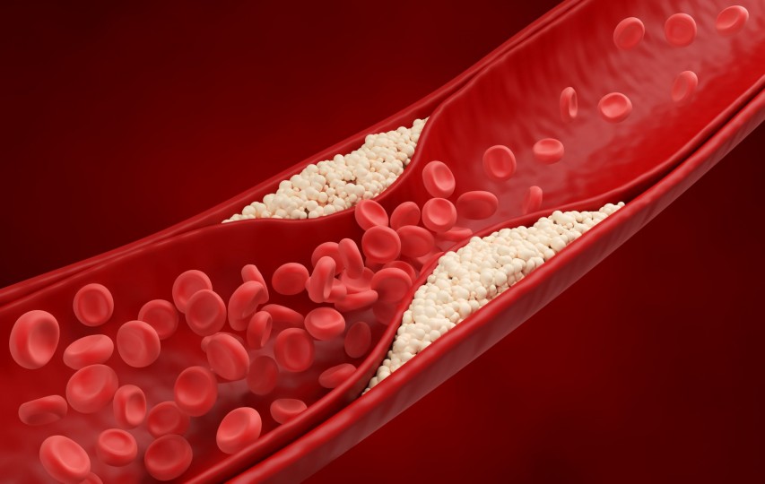
Clogged blood vessels | Photo: Umi No Shizuku Fucoidan (www.kfucoidan.com) |
Vascular Stenosis and the Limitation of Precedent Researches for Early Detection
Vascular Stenosis occurs when internal blood vessel stress increases due to plaque formed by accumulated cholesterol. This condition raises the risk of other health issues, such as neurological diseases or stroke, if the plaque breaks down and travels through the bloodstream.
The mortality risk associated with vascular stenosis has led to various research efforts for detecting the condition in its early stages. For example, studies on constructing blood vessels using a three-dimensional printer, developing a microfluidic method, and applying computational fluid dynamics (CFD) have been conducted to advance the understanding and treatment of the disease.
Although various studies have been conducted, the lack of methods for measuring internal blood vessels and visualizing the results remains a limitation in this field of research. Prof. Park explained that the team aimed to overcome this shortcoming through their research.
PDA Sensor Production and Reaction Mechanism Analysis
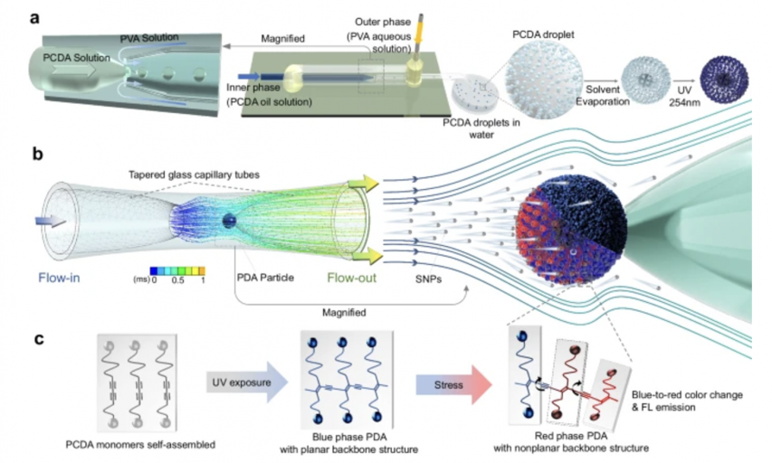
The team created a fluorogenic sensor using PDA particles and analyzed the internal conditions of the vascular stenosis model. Prof. Park explained, “The recent study successfully generated a PDA particle as thick as a human hair, changing its color and emitting fluorescence under mechanical stress. We believed that this feature would be suitable for measuring small areas of the body, such as internal blood vessels.”
The team conducted the study through three stages. In the first stage, they generated the PDA particles at the same scale and examined their reactions. Next, they tested how the sensor responds to various conditions that might occur in the blood vessels, such as fluid viscosity or flow speed. In the final stage, they used CFD to digitize and visualize the data collected by the sensor.
Throughout the study, the team discovered various factors that influence the sensors. The factors included heat, physical impact, flow speed, fluid viscosity, particle size within the fluid, and the duration of stress exposure. They also found that PDA particles emit fluorescence when subjected to these conditions, and they change color from blue to red if enough energy is accumulated from the stress.
Among the conditions, the presence of particles within the fluid had the greatest impact on the sensor. Additionally, the sensor responded most actively when silica nanoparticles, red blood cells, and yeast cells were injected into the vascular stenosis model.
Moreover, the team discovered that the sensor only responds under stress and that the size of the PDA particles is related to the sensor's sensitivity. These observations suggest that exposing the sensor to stress for a certain duration is crucial during measurement and that the sensor’s sensitivity can be adjusted by altering the particle size.
The team utilized the K-omega shear stress transport turbulence model to obtain results. This model is used to predict how the air or fluid moves under specific conditions. Using this approach, they collected digital data and visualized it through CFD, ultimately introducing a new method for measuring stress in internal blood vessels.
Experimental procedure overview | Photo: Nature (www.nature.com) |
Significance and Outlook of the Research
The significance of this study lies in it being the first attempt at using PDA in the vascular field. Prof. Park explained, “PDA particles have been used in various types of sensors. However, using them to investigate stress occurring in small areas of the body, such as the blood vessels, is unprecedented.”
He also underscored overcoming previous challenges: “By measuring the stress occurring in the vascular stenosis model in real-time and visualizing it, we have introduced a new method for monitoring blood vessels. This represents an improvement over previous limitations, which lacked effective monitoring methods.”
Although the study made significant progress with its unprecedented approach, further steps are needed for real-world application in cardiovascular disease checkups. Prof. Park stated that systemic improvements are necessary to advance to the clinical stage. Additionally, further research should be conducted using human body mimicry models to facilitate the sensor in real-world settings.
Conclusion
The team concluded their study with meaningful results. They created a fluorescent sensor using PDA particles and measured the internal condition of the vascular stenosis model. Using this sensor, they successfully collected and visualized digital data. The team’s efforts represented a remarkable attempt and a significant step toward overcoming previous challenges, ultimately suggesting a new monitoring method.
From now on, the team plans to further develop their findings. Prof. Park said, “We are planning a follow-up study to design a more sophisticated vascular mimicry model and enhance the sensitivity of the PDA sensor.”
With the team’s achievements and continued efforts, their work is expected to positively contribute to the prevention of cardiovascular diseases in the future.
There are no registered comments.
- 1
- 2
- 3
- 4
I agree to the collection of personal information.


